
Stress transmission and fracture in collagen networks
Post-doc project
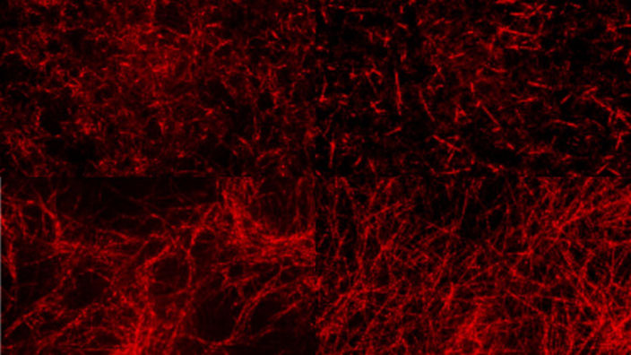
The aim of this project is to tackle the important biophysical and bioengineering issue of understanding the nonlinear mechanical properties of fibrous tissue. Soft tissue connects, surrounds or supports internal organs and bones and exhibits a large variety of architectures1 and mechanical properties2, which vary from organ to organ and according to patho-physiological conditions. Nonlinear phenomena within fibrous networks are known to be crucial for their bulk mechanical properties and for the behaviour of cells within them3, be it physiological (fibroblast tissue maintenance4, immune cell migration5) or pathological (cancer-associated fibroblast remodelling6). While some of these effects such as the buckling/unbuckling of fibers, strain localisation and network connectivity have been shown to play a crucial role in the nonlinear elastic response7-10, the feedback with other effects such as fiber slidings for the stress transmission and for the fracture of these networks remains poorly understood.
The expected breakthrough is to determine and to model the microscopic mechanisms leading to fracture and to long range stress transmission under large deformations in collagen networks. The microstructural properties of these networks suggest that the interplay between fiber sliding, network morphology as well as buckling are key to understand the remodelling and damage11. Extensive coarse-grained molecular dynamics simulations will be used in order to shed light on their specificities while providing a microstructural view of the networks that can be directly compared to experimental visualisation. The project has 3 main steps:
- to establish how the preparation protocols of the networks imply frustrations in the microstructure leading to residual stress,
- to investigate how the network topology, the buckling and sliding of fibers encode the nonlinear response,
- to develop a network model at the mesoscale and in turn a continuum model at the macroscale, allowing to efficiently simulate the material behaviour and to quantitatively compare its properties to those measured by our experimental collaborators.
The combined expertise of 3SR and Liphy laboratories allows to envision this multiscale approach and to expect a breakthrough in the understanding of the nonlinear properties of fibrous networks.
REFERENCES
1 E. Wershof, D. Park, R. P. Jenkins, D. J. Barry, E. Sahai, and P. A. Bates. “Matrix feedback enables diverse higherorder patterning of the extracellular matrix”. en. In: PLOS Computational Biology 15.10 (Oct. 2019). Ed. by P. K. Maini, e1007251. issn: 1553-7358. doi: 10.1371/journal.pcbi.1007251. url: https://dx.plos.org/10.1371/journal. pcbi.1007251
2 I. Levental, P. C. Georges, and P. A. Janmey. “Soft biological materials and their impact on cell function”. en. In: Soft Matter 3.3 (2007), pp. 299–306. issn: 1744-683X, 1744-6848. doi: 10.1039/B610522J. url: http://xlink.rsc.org/?DOI=B610522J
3 A. Erlich, J. Etienne, J. Fouchard, and T. Wyatt. “Review: How dynamic prestress governs the shape of living systems, from the subcellular to tissue scale”. In: Interface Focus (Special issue ‘Complex Rheology in Biological Systems’ 2022). In press. doi: 10.1098/rsfs.2022.0038. url: https://arxiv.org/abs/2209.01440.
4 W. R. Legant, A. Pathak, M. T. Yang, V. S. Deshpande, R. M. McMeeking, and C. S. Chen. “Microfabricated tissue gauges to measure and manipulate forces from 3D microtissues”. en. In: Proceedings of the National Academy of Sciences 106.25 (June 2009), pp. 10097–10102. issn: 0027-8424, 1091-6490. doi: 10 . 1073 / pnas . 0900174106. url: https://pnas.org/doi/full/10.1073/pnas.0900174106
5 S. van Helvert, C. Storm, and P. Friedl. “Mechanoreciprocity in cell migration”. In: Nat Cell Biol 20 (1 2018), pp. 8–20. doi: 10.1038/s41556-017-0012-0.
6 E. Sahai, I. Astsaturov, E. Cukierman, D. G. DeNardo, M. Egeblad, R. M. Evans, D. Fearon, F. R. Greten, S. R. Hingorani, and T. Hunter. “A framework for advancing our understanding of cancer-associated fibroblasts”. In: Nature Reviews Cancer 20.3 (2020). Publisher: Nature Publishing Group, pp. 174–186.
7 P. Ronceray, C. P. Broedersz, and M. Lenz. “Fiber networks amplify active stress”. In: Proc. Natl. Acad. Sci. U.S.A. 113 (11 2016), pp. 2827–2832. doi: 10.1073/pnas.1514208113.
8 C. P. Broedersz, X. Mao, T. C. Lubensky, and F. C. MacKintosh. “Criticality and isostaticity in fibre networks”. In: Nature Physics 7.12 (2011), pp. 983–988.
9 F. Burla, S. Dussi, C. Martinez-Torres, J. Tauber, J. van der Gucht, and G. H. Koenderink. “Connectivity and plasticity determine collagen network fracture”. In: Proceedings of the National Academy of Sciences 117.15 (2020), pp. 8326– 8334.
10 G. Grekas, M. Proestaki, P. Rosakis, J. Notbohm, C. Makridakis, and G. Ravichandran. “Cells exploit a phase transition to mechanically remodel the fibrous extracellular matrix”. In: J. R. Soc. Interface. 18 (175 2021), p. 213. doi: 10.1098/ rsif.2020.0823.
11 V. R. Sherman, W. Yang, and M. A. Meyers. “The materials science of collagen”. In: Journal of the mechanical behavior of biomedical materials 52 (2015), pp. 22–50.


