
Margination phenomena: segregation in blood and model suspensions
Post-doc project
Living matter offers the opportunity to study the behaviour of original phenomena in which some physical mechanisms prevail and remain to elucidate. In that perspective, blood is a suspension of elastic objects, namely Red Blood Cells (RBCs), whose large deformability in the vessels leads to rheological properties of complex fluids and to intriguing structuration phenomena such as margination. Margination consists in a segregation of blood cells under flow: RBCs are located at the centre of the vessel while White Blood Cells (WBCs) and platelets are at the periphery close to the endothelial cells covering the vessel. Margination has a strong impact on the physiological response as platelets are close to the vessel wall in the case of injury and WBCs only need to migrate through the endothelial barrier to reach infected tissues. While known from many decades in medecine, the detailed mechanisms and modelling of margination are still a challenge despite the numerous numerical investigations and the interest in taking advantage of it for separation purposes.
Recently, there has been a growing international interest of numerical/theoretical teams, who have proposed different scenarios. All these results tend to focus on the major role played by the difference of rigidities between healthy RBCs, platelets and WBCs generating negative and positive migrations normal to the direction of the flow.
However there is an obvious lack of controlled experiments in microfluidic model systems to provide quantitative data for a deeper understanding of margination and also to highlight non trivial effects that preliminary experiments have revealed.
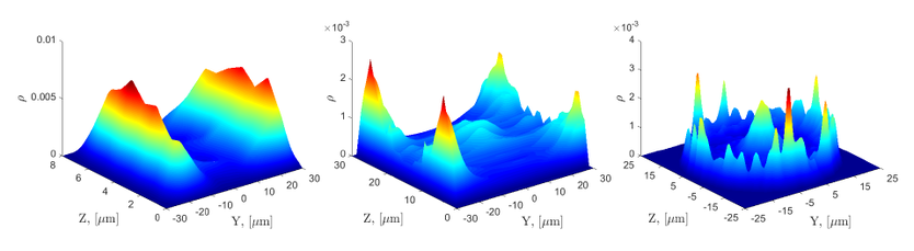
Drawing on our experience, we propose an experimental investigation of margination phenomena with a simplified blood model containing two populations of RBCs, namely healthy and rigidified by glutaraldehyde. The suspensions containing a fraction of rigidified RBCs will flow in straight microfluidic channels at different flow rates, hematocrit and sections. The distribution of rigidified RBCs in the section will be determined by fluorescence and confocal microscopy. We will also study the evolution of margination with the distance from the channel input. Preliminary experiments show tremendous and unexpected effects of the channel geometry (section): square, circle and high aspect ratio rectangle (see figure).
Figure caption: Effect of channel geometry on margination (from Chachanidze et al. 2017). Left – channel with high aspect ratio 8/60 = depth/width, distances in micrometers. The concentration of rigidified RBCs is high at the two channel sides. Middle – channel with aspect ratio 30/60. Margination is preferentially localized at the four corners with a secondary peak at the center. Right – tube channel with 50 µm of diameter. Margination is localized at the wall and at the center. These differences of spatial distributions have never been observed experimentally. Note that the central secondary peak has only been observed in 2017 by Qi et al. in one numerical simulation in square geometry.

CONTACTS
- PI: Clément de Loubens
- Co-PI: Thomas Podgorski
- Post-doc: Revaz Chachanidze

PARTNERS
- LRP
- LIPhy

FUNDING
Tec21