
Results published in 2018 in Scientific Reports
Researchers from the Laboratoire 3SR and their colleagues have released the first 3D high-resolution images of the human vocal folds thanks to X-ray microtomography coupled with a phase retrieval technique. These images reveal the complex structure of larynx tissues and open the way towards a better understanding of their virbo-mechanical properties.
Human vocal folds possess the outstanding ability to endure large but reversible deformations and to reach vibration frequencies between 50 and 1 500 cycles per second. This unique performance mainly results from their complex specific 3D and multiscale structure. The characterisation of each element of this structure remains a great challenge today, even with the use of imaging techniques such as confocal microscopy (limited depth of field), magnetic resonance (limited spatial resolution) or X-ray absorption microtomography (limited phase contrast).
X-ray tomography for a multiscale 3D analysis
The researchers from the Laboratoire 3SR and their colleagues (1) have used high-resolution synchrotron X-ray microtomography associated to phase retrieval. Thanks to this technique, they have been able to report the first ex vivo 3D images of human vocal-fold tissues at multiple scales.
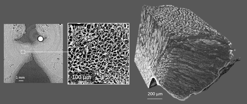
Various relevant descriptors of the structure were extracted from the images: the geometry of vocal folds at rest or in a stretched phonatory-like position, the shape and size of their layered fibrous architectures, the orientation, shape and size of the muscle fibres as well as the set of collagen and elastin fibre bundles constituting these layers.
The experimental methods used in this work open a promising way towards a better understanding of the micromechanics of the vocal folds, their deformation, and the link between the fibrous architecture and the vibratory properties. This work should provide valuable guidelines for the design of new mimetic biomaterials for the next generation of artificial larynges.
(1) GIPSA-lab, LADAF, NOVITOM, ESRF
Figure caption
Multiscale X-ray tomographic images of a human vocal fold. Left: global view. Center: detail of the tissue. Right: 3D reconstruction of the network of muscular, collagen and elastic fibres (adapted from Bailly et al., in Scientific Reports, 2018)
Reference
Bailly L, Cochereau T, Orgéas L, Henrich Bernardoni N, Rolland du Roscoat S, McLeer-Florin A, Robert Y, Laval X, Laurencin T, Chaffanjon P, Fayard B and Boller E: 3D multiscale imaging of human vocal folds using synchrotron X-ray microtomography in phase retrieval mode. Scientific Reports, Nature Publishing Group, 2018, 8 (1).
CONTACTS


LABORATORY
Laboratoire sols solides structures et risques
FOR MORE INFO
View the project 