
Results published in 2019 in Microvascular Research
Researchers from the Interdisciplinary Laboratory of Physics have determined a scaling law for the transversal drift of red blood cells in blood vessels.
Blood is made of an assembly of cells suspended in the plasma. From a mechanical point of view, blood is therefore considered as a suspension of deformable vesicles (red blood cells) spread in a liquid and circulating in the complex network of vessels and capillaries, similarly to a crowd walking in a labyrinth of corridors. And just like humans do, red blood cells tend to keep away from the walls and to migrate towards the centre of the channel. When reaching an intersection, this migration behaviour plays an important role in the distribution of red blood cells in each branch: which direction is a cell going to choose? What is a cell’s trajectory going to be depending on its distance to the centre of the blood vessel? How long does it take for a cell to get back again to the centre of the flow after a turn? And what if a cell has not reached its equilibrium cruising position before the next intersection?
An ingenious set up to study the drift of individual cells
To answer these questions, the researchers from the Interdisciplinary Laboratory of Physics have designed an ingenious microfluidic “T” shaped experimental set up, to reproduce the hydrodynamics of blood vessels and analyse the trajectories of individual red blood cells under controlled conditions. Using dilute suspensions of cells, and altering their mechanical properties by artificially tuning their rigidity or selecting populations of various ages, they were able to work out a scaling law for the transverse migration velocity of red blood cells in channels. This “drift law” proved valid in a large range of parameters and lays a solid basis for the understanding of the microcirculation. Such results are also applicable to a number of practical situations, including microfluidic systems and biomedical devices.
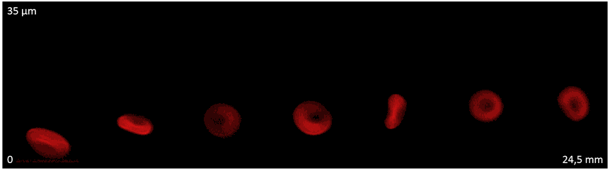
Figure caption
Representation of the trajectory of a red blood cell in a 34.8 µm wide microfluidic channel. The images were taken from 7 windows of size L=350 μm at different positions in the channel. About 200
red blood cells were observed in each window and the positions shown on the figure were chosen to represent the median observed trajectory (adapted from Losserand et al., in Microvascular
Research, 2019)
Reference
S. Losserand, G. Coupier & T. Podgorski. 2019. Migration velocity of red blood cells in microchannels. Microvascular Research, 124: 30-36.
CONTACTS


Gwennou Coupier
LABORATORY
LaboratoireInterdisciplinaire de Physique

FOR MORE INFO
Read the full article online 