
PLATFORMS
PLATFORMS
» Confocal microscopy

Tec21 has acquired the latest version of Leica's confocal microscope: the TCS SP8
This high performance laser fluorescence microscope was delivered and installed at the Dyfcom Department of the LIPhy (Laboratory for Interdisciplinary Physics) in December 2012, where 5 training sessions were organised, and attended by about twenty researchers.
Confocal microscopy is a fluorescence optical imaging technique based on point illumination, which eliminates out-of-focus light, giving highly-resolved focal planes and high-contrast images. The scanning of a sample along the z-axis (vertical dimension) enables the reconstruction of a three-dimensional image through the stacking of all focal planes.
The TCS SP8 was chosen for its versatility: it has 7 possible excitation wavelengths (from 458 to 633 nm), and a fully customizable light detection system, allowing the user to monitor virtually any 3 different fluorophores simultaneously on a given specimen. Moreover, the SP8 high-speed imaging resonant-scanner system can process up to 29 images per second (512x512 pixels).
Such features enable multi-mode 3D imaging of specimens (3 wavelengths, [x, y, z] scan) as well as 2D or even 3D video captures ([x, y, t] or [x, y, z, t] scans).
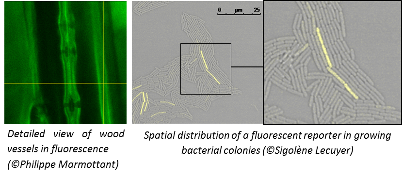
Our confocal microscope is currently used in different research programs, studying for instance biofilm behavior under fluid mechanical constraints, sap cavitation phenomenon in wood vessels, or the structure of bones.
Tec 21 is already considering the upgrade of this system with an automated motorised stage as well as an environmental chamber. The latter will provide control over the temperature, relative humidity, and CO2 concentration, thus significantly widening the application fields for the microscope. The chamber should be added within a few months.
For all technical information and support, please contact Sigolène Lecuyer.
An automated motorised stage installed on the confocal microscope: new perspectives in research
The confocal microscope was recently upgraded with an automated motorised stage raising great perspectives in terms of the use of the microscope.
After setting the [x, y, z] coordinates of different spots of interest in the sample, the operator can start a scanning sequence during which the microscope will automatically move the stage and acquire images of each of the registered spots over a period of time and with a frequency to be defined by the operator. A virtually unlimited number of spots can therefore be monitored in the same sample over long-time experiments.
Such a device enables to cope with one of the main obstacles encountered when studying biological objects, i.e. variability. The following illustration is a prime example.
Researchers from the Laboratory for Interdisciplinary Physics who study the process of biofilm production in a model bacteria, have been able to survey the growth of more than 20 different individuals on the same plate, from the one-cell-stage to the colony, over a night period. The microscope acquired an image of each of the target bacteria every 10 minutes (the video below shows examples of growth patterns observed on 4 different colonies during this experiment).
The scientists have therefore been able to collect data on more than 20 replicates during the same experiment. Without the automated motorised stage, the acquisition of such a data set would have required… 20 nights!
Other custom modes enable the wide field imaging of a sample through automated grid scanning procedures (see animation).
For more information about confocal microscopy, please contact Sigolène Lecuyer.
<< back